Background
DNA damage is a key driver of cell dysfunction, contributing to processes like cellular aging and apoptosis. Although DNA damage is constantly challenged by endogenous factors, it is also vulnerable to exogenous stressors. Exogenous stressors include UV light, chemotherapy, and reactive oxygen species, as well as physical stresses like osmotic pressure and mechanical stimuli. These physical stresses are present daily for epithelial cells, which line the skin, blood vessels, lungs, and digestive tract. For instance, alveolar epithelial cells undergo cyclic strains of 4–12% during normal breathing, intestinal epithelial cells experience continuous deformation during peristalsis, and our skin can stretch by as much as 25% during everyday joint movements and sports.
Although numerous studies have explored how mechanical forces and osmotic pressure influence epithelial cells, a fundamental question remains unanswered: how do epithelial cells protect themselves, and ultimately prevent DNA damage, from the mechanical forces and osmotic stress they experience daily? The recent study by Yang and colleagues titled “Transient perinuclear actin rings prevent cell aging and apoptosis via nuclear mechanical protection,” published in the July 07, 2026, issue of Biophysical Journal, addresses this question by uncovering an actin-based mechanism that mechanically protects the nucleus during acute stress.
Approach
Yang et al. postulate that epithelial cells may possess a mechanical protection mechanism relying on assembly of a perinuclear actin cytoskeleton and investigate this via experiments and simulations. For their primary in vitro model, they used Madin-Darby canine kidney (MDCK) epithelial cells, with additional validation in MDA-MB-231 breast cancer cells and mouse embryos. To investigate how epithelial cells protect their nuclei from mechanical stress, the authors subjected cells to hypertonic and hypotonic shock, as well as localized mechanical compression by using atomic force microscopy (AFM). By combining AFM with fluorescence and confocal microscopy, they monitored changes in actin organization and nuclear morphology in real time, while also quantifying mechanical properties of the nucleus. To determine whether these cytoskeletal changes were functionally important, the authors perturbed key components of the cell mechanical response, including inhibiting Arp2/3-mediated and formin-mediated actin polymerization, stabilizing actin filaments to prevent actin depolymerization, disrupting actin ring assembly through calcium chelation, and knocking down Lamin A/C to weaken nuclear mechanical integrity. Complementing the experimental work, Yang and colleagues developed a computational model based on their experimental observations and previous studies, enabling them to gain mechanistic insight into the cellular responses to mechanical stress.
Key findings
Mechanical stress triggers transient perinuclear actin rings
The authors first tested how cells respond to rapid mechanical perturbations by exposing MDCK cells expressing fluorescently labeled actin (LifeAct-EGFP) to hypotonic shock. They observed a transient ring of actin assembled around the nucleus, which then disappeared within 30 min from the shock. Ring formation was dictated by the amplitude and rate of osmotic pressure decreases: increasing the hypotonic shock from small to large amplitude increased the proportion of cells forming an actin ring from ~20% to nearly 80% while reducing the time required to form a ring from ~12 to just 3 minutes. In contrast, neither a gradual decrease in osmolarity nor hypertonic shock induced perinuclear actin ring formation, with the same transient response also observed in mouse embryos. Because hypotonic shock increases cortical tension via cell swelling, the authors asked whether direct mechanical loading could elicit the same response. Using AFM to apply controlled compression, they found that only rapid loading or unloading led to perinuclear actin ring formation, reinforcing the finding that epithelial cells reorganize their actin cytoskeleton only under rapid and significant mechanical perturbations.
Calcium signaling and myosin contractility in perinuclear actin ring dynamics
Yang et al. next assessed whether calcium signaling mediates the observed response to mechanical forces. Using a fluorescent calcium indicator, they found that intracellular Ca2+ concentration increased immediately after both hypotonic shocks and AFM indentation, but not hypertonic shock, with Ca2+ signal peaking consistently before perinuclear actin ring formation, suggesting that calcium signaling initiates actin reorganization. Experiments confirmed this hypothesis: chelating extracellular Ca2+ with EDTA abolished perinuclear actin ring formation, although increasing Ca2+ influx with the calcium ionophore A23187 rapidly induced it. Yang et al. also identified TRPV4 as the upstream mediator of this response. Furthermore, the authors proposed that calcium-mediated actin depolymerization transiently increases the intracellular pool of G-actin for cytoskeletal remodeling. Consistent with this idea, blocking either actin depolymerization (Jasplakinolide), actin polymerization (Latrunculin A), or myosin contractility (Blebbistatin/Rhosin) abolished perinuclear actin ring formation despite calcium influx, indicating that both myosin-driven contraction and actin turnover are required for perinuclear actin ring assembly.
Modeling perinuclear actin dynamics
On the basis of their experimental observations and previous studies, Yang and colleagues developed a computational model to gain further mechanistic insight into perinuclear actin dynamics. The two-dimensional model incorporated actin polymerization, actin depolymerization, myosin-driven contraction, and monomeric actin diffusion, while treating the nucleus as a rigid spatial geometric obstacle to simplify calculations. In agreement with their experimental observations, simulated mechanical stress triggered calcium-dependent cortical actin depolymerization, increasing the local pool of G-actin transiently. As G-actin diffused toward the nucleus, it repolymerized into F-actin, with myosin leading to further network contraction. Because contraction of the F-actin network was blocked by the nucleus, a transient perinuclear actin ring appeared in simulations, recapitulating experiments.
Arp2/3 drives actin ring assembly
To identify the molecular machinery driving actin ring formation, the authors combined simulations and experiments. Simulations predicted that inhibiting formins would not affect perinuclear actin ring formation, a finding confirmed experimentally with formin inhibitor SMIFH2. In contrast, perinuclear actin ring assembly vanished when inhibiting the Arp2/3 complex, regardless of osmotic shock or mechanical compression. The computational model also predicted that actin rings are not assembled specifically around the nucleus through LINC complex-mediated interactions, but instead around any intracellular obstacle. When introducing microbeads into cells, Yang et al. demonstrated that perinuclear actin ring formation is not a nucleus-specific process, but instead a nonspecific adaptive response of the actin cytoskeleton to intracellular obstacles during acute mechanical stress.
Perinuclear actin rings transiently stiffen the nucleus
By using AFM indentation, Yang and colleagues observed transient stiffening of the nucleus during actin ring assembly, prompting them to investigate the source of this mechanical response. Although total Lamin A/C levels remained unchanged, the authors observed enrichment of Lamin A/C at the nuclear envelope specifically during actin ring assembly, indicating that Lamin A/C redistribution contributes to nuclear stiffening. Interestingly, knocking down Lamin A/C by over 90% only partially reduced the magnitude of nuclear stiffening (40% to 18%), indicating that Lamin A/C could not explain this response. The authors proposed that mechanical confinement from the perinuclear actin ring may also contribute to the nuclear stiffening. Their simulations supported this idea, predicting that the myosin-mediated contraction of the actin ring generates compressive stresses sufficient to stiffen the nucleus. Thus, the observed transient nuclear stiffening probably arises from the combined effect of Lamin A/C redistribution and mechanical confinement by the perinuclear actin ring.
Perinuclear actin ring formation protects the nucleus
To determine whether the transient actin ring serves a functional role in regulating nuclear morphology and stability, Yang and colleagues combined 3D reconstructions and finite element method simulations. They found that the perinuclear actin ring mechanically confines the nucleus, limiting its deformation during hypotonic cell swelling. As a result, nuclei surrounded by an intact actin ring had lower nuclear envelope tension despite continued swelling. Moreover, actin ring assembly also reduced the incidence of nuclear envelope rupture, demonstrating that it acts as a mechanical safeguard during acute external mechanical stresses.
Protection from DNA damage, senescence, and apoptosis
Having established that the perinuclear actin ring mechanically safeguards the nucleus, Yang and colleagues tested whether this protection translates into reduced DNA damage. By using a comet assay and γH2AX staining (a canonical marker of DNA double-strand breaks), Yang and colleagues observed significantly greater DNA damage when actin ring assembly was inhibited under hypotonic shock, indicating that actin ring–mediated compressive stress can directly mitigate DNA damage during mechanical stress. Finally, the authors asked whether this mechanical protection extends beyond DNA damage to long-term cell outcomes like senescence and apoptosis. By quantifying markers of cell aging and apoptosis during hypotonic shock in control cells versus cells unable to form the perinuclear actin ring, Yang et al. demonstrated that actin ring assembly not only prevents DNA damage but also protects cells from the cumulative consequences of repeated mechanical stress, including premature senescence and apoptosis.
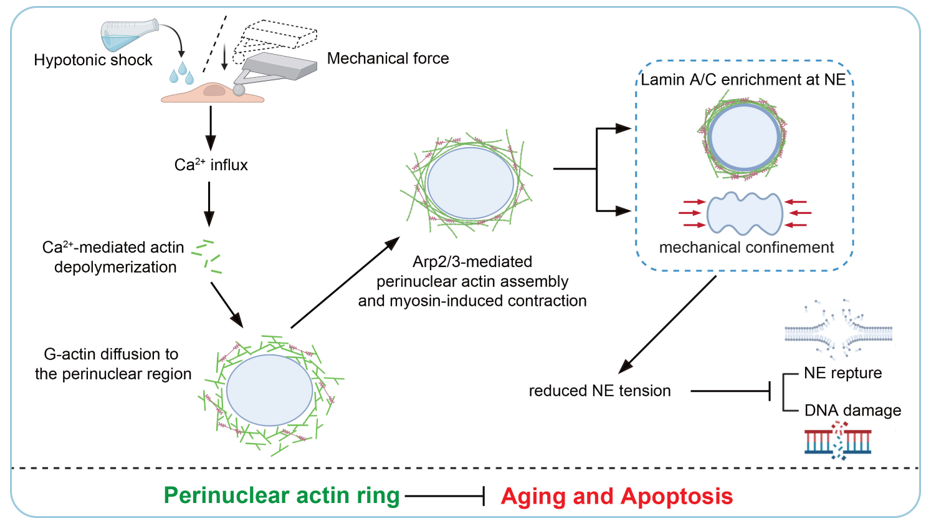
Figure 1: Schematic summarizing the mechanism proposed by Yang et al. Acute mechanical stress (e.g., hypotonic shock or mechanical compression) triggers calcium influx, leading to calcium-mediated actin depolymerization, G-actin redistribution to the perinuclear region, and Arp2/3-dependent assembly of a transient perinuclear actin ring. This results in Lamin A/C enrichment at the nuclear envelope (NE) and mechanical confinement and stiffening of the nucleus, reducing NE tension and preventing NE rupture and, in turn, protecting cells from DNA damage, cell aging, and apoptosis. Figure from Yang et al. 2026.
Conclusions
The current study by Yang and colleagues combines live-cell imaging, biophysical measurements, computational modeling, and pharmacological perturbations to uncover a mechanical defense mechanism that protects epithelial cells, and in particular, their nuclei, from acute physical stress. Importantly, this protective mechanism appears not to be unique to the nucleus but instead represents a general adaptive response of the actin cytoskeleton capable of providing transient mechanical support in response to intense external stimuli. By mechanically safeguarding the nucleus, this transient reorganization of the intracellular actin network ultimately prevents DNA damage, senescence, and apoptosis.
— Ilaria Di Meglio