A highly sensitive new blood test can detect rare cancer proteins
Researchers at Johns Hopkins University developed a new blood test that can identify proteins-of-interest down to the sub-femtomolar range with minimal errors
Baltimore, MD – Proteins that normally reside inside cell nuclei have never been found in the blood, until now. A new blood test developed at the Johns Hopkins University by Shih-Chin Wang and Chih-Ping Mao—graduate students in Jie Xiao’s lab in the Department of Biophysics and Chien-Fu Hung’s lab in the Department of Pathology—can identify individual molecules in human blood samples with minimal detection errors. Among the molecules that they used their new test to find was a mutated protein thought to be restricted to the inside of cells, mostly within the nucleus. It is the first time that single-molecule imaging has been applied to visualize disease-causing molecules in blood. They will present their research at the 63rd Biophysical Society Annual Meeting, to be held March 2 - 6, 2019 in Baltimore, Maryland.
Wang and colleagues call their new approach Single-Molecule Augmented Capture (SMAC). They used this new technique to detect molecules commonly screened for in standard blood tests, like prostate-specific antigen. And they were also able to detect rare intracellular proteins, secreted proteins and membrane proteins, including the cancer-associated proteins mutant p53, anti-p53 autoantibodies and programmed death-ligand 1 (PD-L1).
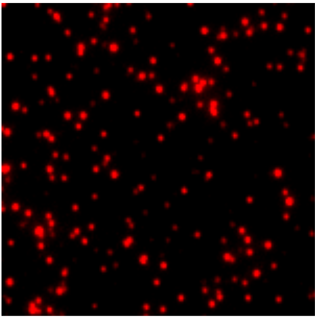
Mutant p53 is a well-known tumor-specific nuclear protein and has never before been detected in the blood, likely because current tests cannot detect its extremely low blood concentrations. Wang and colleagues found mutant p53 or anti-p53 autoantibodies in samples from patients with ovarian cancer, but not in patients without cancer. PD-L1 is also found on the surface of some cancer cells and has recently been effectively targeted with immunotherapy to combat cancer. Knowing whether or not a patient’s tumor expresses PD-L1 is a crucial first step in this treatment—and SMAC may be able to identify cancers that have PD-L1 at low levels that are undetectable by standard blood tests.
“With SMAC, we have brought single-molecule imaging into the clinical arena. By visualizing and examining individual molecules released from diseased cells into the blood, we aim to detect diseases more accurately and gain new insights into their mechanisms,” Mao said.
Wang and colleagues are hopeful that their test will one day be commercially available.
###
Caption: This SMAC image shows mutant p53 proteins in the blood of an ovarian cancer patient. Individual clusters of p53 molecules are represented by red spots. Image courtesy of Shih-Chin Wang.
