High fat diet causes thickening of arteries down to the cellular level
Researchers at the University of Illinois show that the membranes of cells surrounding arteries get stiffer and thicker in response to a high fat diet, due to both LDLs and oxidized LDLs
BALTIMORE, MD – Atherosclerosis is a tough problem—arteries get thicker and stiffer, which can lead to heart disease and stroke, but it is not known precisely how cholesterol causes this thickening. Cholesterol is a tiny fat molecule that circulates in our blood stream with the help of lipoproteins. High levels of low-density lipoproteins (LDLs) in the blood is a key risk factor for atherosclerosis. And a variant of LDLs, called oxidized LDLs, may also be contributing to arterial plaques. Manuela Ayee, who worked with Irena Levitan at the University of Illinois, will present their research on how those two LDLs cause thickening at the cellular level at the 63rd Biophysical Society Annual Meeting, to be held March 2 - 6, 2019 in Baltimore, Maryland.
Cholesterol is not all bad, it’s an essential fat that cells need to make membranes and steroid hormones. When it is transported through our bodies, it needs a lipoprotein carrier. LDLs carry cholesterol away from the liver to cells, and high-density lipoproteins (HDLs) return cholesterol to the liver. LDL has long been believed to be the cause of atherosclerosis, but recent evidence suggests that oxidized LDL is also a key player. Ayee and Levitan wanted to know if it is one, or both, of these LDLs that is the main problem.
Ayee and Levitan fed mice either a normal well-balanced diet, or “western high fat diet” that was developed to mirror the levels of fat, protein, and carbohydrate typically found on a fast food menu. They observed that the mice consuming the latter diet quickly developed stiffer arteries, which occurred down to the layer of endothelial cells surrounding the blood vessels. They measured the levels of LDLs and oxidized LDLs in these mice so they could apply the same concentrations to human endothelial cells in culture, then did precise measurements using atomic force microscopy of the membrane tension and the stiffness of the cytoskeleton.
In culture, compared to cells without added lipoproteins, physiological levels of LDLs and oxidized LDLs each caused cell membrane thickening and increased tension when added individually, and together the change was amplified.
“To our surprise, a very small amount of oxidized LDL dramatically changes the structure of the cell membrane for the worse,” Ayee said.
Their results suggest that changes in blood lipid levels due to diet can fundamentally disrupt the integrity of the endothelial cell layer.
“We think that the changes at the cell membrane level may allow the processes involved in atherosclerosis to begin,” Ayee concluded.
###
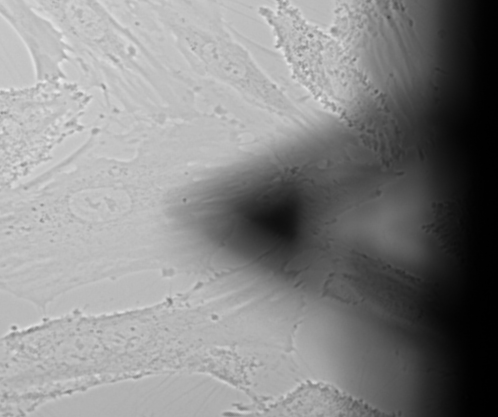
A human aortic endothelial cell being probed by an atomic force microscope (the probe is the triangle). Image courtesy of Manuela Ayee.